Ultrasound of the Feet?
February, 2019
Cyrus Khorrami, MD
Radiologist, Medical Director
Ultrasound imaging is a very common way to image the inside of the body. It is a quick and painless test that uses high-frequency sound waves to create pictures of the body. It uses no radiation, which makes it safe enough to use on pregnant women and their unborn babies. Ultrasound technology is also used to image the organs of the abdomen and pelvis as well as to give a clear picture of the blood vessels. More frequently, we are using ultrasounds to take images of the feet.
Q: What can the ultrasound show us in the feet?
A: The ultrasound is excellent at imaging the tendons of the feet. Tendons are in the ends of muscles that attach to the bone. The tendons surround the foot and this allows us to move the foot in every direction. The tendons can get damaged and cause foot pain. Sometimes surgery needs to be performed to correct a torn tendon. Ultrasound is also commonly used to image the plantar fascia. The plantar fascia is fibrous tissue on the bottom of the foot that connects the heel bone to the toes. The plantar fascia can get inflamed, causing pain usually at the heel. This is called plantar fasciitis. Ultrasound can detect plantar fasciitis and its level of severity. Ultrasound is a great tool at imaging the Achilles tendon (also called the Achilles heel). This large tendon is located at the back of your heel and attaches to the bone in the back of your foot. It can become inflamed and painful (called Achilles tendinitis). In some cases, the Achilles tendon can tear. Depending on how badly the tendon is ruptured, it may require surgery to repair.
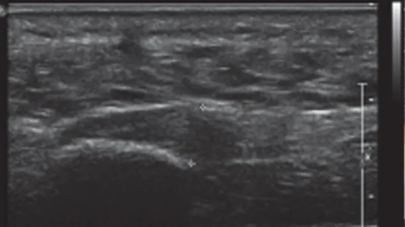
Q: What else besides tendons can the ultrasound image?
A: Anything abnormal in the soft tissue can be seen by ultrasound. It can be used to detect collections of fluid, such as pools of blood from bruising or an abscess from an infection. There is a condition called Morton’s neuroma. This occurs when there is thickening of tissue around the nerves. This is located at the balls of your feet, usually at the level of the third and fourth toes. This can be very painful. Many patients describe the sensation of having a rock in their shoe. An ultrasound examination can usually find a Morton’s neuroma and help guide the surgeon to remove it.
Q: Can the ultrasound see the bone of the foot? Can it see arthritis?
A: Unfortunately, ultrasound is limited in imaging bones. The dense calcium of bone blocks the sounds waves and we only see a shadow. That is why a foot ultrasound is often interpreted with a foot x-ray.
Q: How is the test performed?
A: The shoes and socks are removed. The patient is then asked to either lie down on a table or sit with both feet dangling off the table. Warm gel is applied to the foot and the technologist uses the ultrasound probe to take pictures of the area of concern.